


Columbia Blood Agar, with Hemin and Vitamin K₁ | For 5L
- Product Code: GMNB-CAB11
- Availability: In Stock
Tags: Columbia Agar
Overview
The GMExpression formulation uses the Ellner (1966) Columbia agar base, prepared with Australian DAFF EX188M biosecurity-certified defibrinated sheep blood. The medium permits reading of three independent phenotypes on a single plate: (1) colonial morphology (sub-millimetre Bacteroides & Prevotella through 2–3 mm Clostridium colonies); (2) haemolysis pattern (β-haemolysis of Streptococcus spp., α-haemolysis of viridans streptococci, and the pathognomonic double-zone β-haemolysis of Clostridium perfringens); and (3) pigment production (the bronze-to-black colonies of Prevotella melaninogenica and Porphyromonas gingivalis, the brick-red autofluorescence of Bacteroides ureolyticus group, the chartreuse fluorescence of Veillonella). It differentiates from the existing GMExpression Brucella Agar product line by Columbia base composition (Columbia is richer in beef-heart infusion and peptone), giving more luxuriant colony growth.
We also have
YCFA Modified Medium · YCFA Full Recipe · YCFA Base · Modified Chopped Meat Broth (ATCC 1490) · Chopped Meat with Carbohydrates · Beef Granules
Package Contents
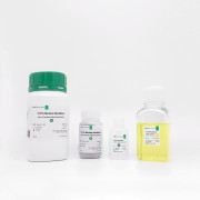
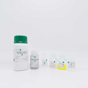
Each GMExpression Anaerobic CBA kit contains:
- Mixture Base — pre-weighed Columbia base + agar (Pantothene-Casein Peptone, Bio-Polytone, beef heart infusion, corn starch, NaCl, agar 13 g/L) for 5 L final volume.
- Stock H — Hemin stock, 25 mg in 5 mL of 0.05 M NaOH, sealed amber vial.
- Stock K — Vitamin K1 stock, 50 mg in 5 mL of 95 % ethanol, amber vial, light-protected.
- Sheep blood pack — 250 mL defibrinated sheep blood (DAFF EX188M certified), shipped at 2–8 °C separately.
- 50 × Petri plates (90 mm) or 100 × Petri plates (60 mm) if pre-poured option selected.
- Instruction manual (A5 booklet, v1.0) with phenotype-reading colour atlas annex.
Customisation options on request: selective variants (kanamycin-vancomycin laked blood = KVLB; Bacteroides-Bile-Esculin = BBE — see related product GMNB-BBE01/KVLB01); chocolatised variant (haemolysed at 80 °C for Haemophilus); 5 % horse blood (in place of sheep); supply as pre-poured plates or as bulk powder + blood for in-house pouring.
Composition — per 1 L equivalent unless stated otherwise
Columbia Agar Base (Ellner 1966; BD Difco 211125 reference; per 1 L)
| Component | Concentration | Function |
|---|---|---|
| Pantothene-Casein Peptone (pancreatic digest of casein) | 10.0 g | High-quality peptide nitrogen source |
| Bio-Polytone (peptic digest of animal tissue) | 5.0 g | Supplementary peptide source |
| Yeast extract | 5.0 g | B-vitamins, growth factors |
| Beef heart infusion solids | 3.0 g | Amino acids, haematin precursors |
| Maize starch | 1.0 g | Trace-toxin binding (absorbs autoclave-derived inhibitors) |
| Sodium chloride (NaCl) | 5.0 g | Osmotic balance |
| Agar (microbiological grade) | 15.0 g | Solidifying agent (note: Columbia base uses 13 g/L, not the more usual 15 g/L) |
Anaerobic supplements (post-autoclave additions)
| Component | Final concentration | Stock and addition |
|---|---|---|
| Hemin (CAS 16009-13-5) | 5.0 mg/L | 5 mL of 1 mg/mL stock (Stock H) per litre of base |
| Vitamin K1 (phylloquinone, CAS 84-80-0) | 1.0 mg/L | 2 mL of 0.5 mg/mL stock (Stock K) per litre of base |
| Defibrinated sheep blood (DAFF EX188M) | 5 % v/v (50 mL/L) | Add at 48–52 °C post-autoclave; pour plates immediately |
Pre-autoclaving pH: 7.3 ± 0.1 at 25 °C. Adjust with 1 M NaOH or 1 M HCl.
Use and Applications
- Primary isolation of obligate anaerobes from clinical specimens — the most-used anaerobic primary plate globally; abscess aspirate, deep-tissue and surgical specimens, blood-culture subculture, intra-abdominal fluid, OB-GYN specimens.
- Differential plate reading haemolysis and pigment phenotypes alongside colonial morphology — C. perfringens double-zone β-haemolysis is essentially pathognomonic; Prevotella / Porphyromonas bronze-to-black pigmentation enables genus-level presumptive identification at 24–48 h.
- UV-fluorescence reading under 365 nm Wood's lamp: brick-red fluorescence (Bacteroides ureolyticus group), chartreuse / lime-green (Veillonella), coral-pink (Porphyromonas asaccharolytica).
- Sub-culture plate for clinical anaerobes after enrichment in BHI-S or Modified Chopped Meat Broth.
- Base for selective derivatives — KVLB (kanamycin + vancomycin + laked sheep blood, for Gram-negative anaerobes), BBE (with oxgall + esculin, for B. fragilis group), PEA (phenylethyl alcohol, for Gram-positive anaerobes), CCMB (cycloserine-cefoxitin-mannitol, for C. difficile).
- Compatible with anaerobic gas-jar / AAE / GasPak workflows at 35–37 °C; reading at 24, 48, and 72 h.
Compatible Microorganisms
Obligate anaerobes
- Bacteroides fragilis ATCC 25285, B. thetaiotaomicron ATCC 29148/29741, B. vulgatus, B. uniformis — grey-white, 2–4 mm, non-haemolytic to weakly α-haemolytic colonies
- Prevotella melaninogenica (ATCC 25845) — small (1–2 mm) colonies developing bronze-to-black pigment by 72 h
- Porphyromonas gingivalis (ATCC 33277) — slower-growing (5–7 d), black pigmented; brick-red Wood's-lamp fluorescence
- Fusobacterium nucleatum (ATCC 25586) — irregular small colonies with breadcrumb morphology
- Clostridium perfringens (ATCC 13124) — large (3–5 mm) colonies with characteristic double-zone β-haemolysis (inner θ-toxin lytic zone, outer α-toxin partial-lytic zone) — essentially pathognomonic
- C. sporogenes (ATCC 19404), C. septicum, C. novyi, C. tetani, C. bifermentans
- Clostridioides difficile (ATCC 9689) — flat irregular ground-glass colonies
- Peptostreptococcus anaerobius, Finegoldia magna (formerly Peptostreptococcus magnus)
- Cutibacterium acnes (formerly Propionibacterium acnes; Scholz & Kilian 2016) — small white colonies on extended incubation
- Veillonella parvula — small grey colonies with chartreuse fluorescence under Wood's lamp
- Actinomyces israelii, Actinomyces meyeri — molar-tooth or breadcrumb morphology, requires 5–10 d incubation
Facultative anaerobes (read alongside)
- Staphylococcus aureus, Streptococcus spp., Enterococcus spp., E. coli, Klebsiella spp. — grow well; useful as positive controls.
Preparation
Critical control points
- Blood-addition temperature. The agar base must be at 48–52 °C when blood is added. Outside this range either chocolatisation (> 55 °C) or premature gelling (< 45 °C) destroys the plate's utility.
- Sheep blood lot QC. Each new blood lot should be tested with C. perfringens ATCC 13124 and S. aureus ATCC 25923 to verify the double-zone β-haemolysis and β-haemolysis patterns respectively before clinical use.
- Wood's-lamp pigment reading. Read pigment fluorescence at 24, 48, and 72 h — Prevotella bronze pigment can take 5–7 d to develop fully; do not read pigment under fluorescent room-light alone, use a 365 nm UV (Wood's) lamp in a dark room.
Cautions
Storage and Expiry · Safety
- Dehydrated powder (Mixture A): 15–30 °C in original packaging. Shelf life 36 months.
- Stock H (hemin): 4 °C, protected from light. Stable 8 weeks.
- Stock K (Vitamin K1): 4 °C, protected from light. Stable 6 months.
- Defibrinated sheep blood: 2–8 °C. Shelf life 28 days from collection.
- Prepared plates, sealed in bags at 4 °C: 4 weeks routine; 6 weeks if double-bagged.
- PRAS plates, vacuum-sealed in AAE + oxygen absorber: 3 months for primary-isolation use.
Safety notes. Defibrinated sheep blood is a BSL-2 biological. Handle in a Class II biosafety cabinet, particularly when preparing plates for culturing BSL-3 pathogens. Hemin in alkaline NaOH is corrosive; pipette with care. Vitamin K1 ethanolic stock is flammable. SDS available on request.
References
- Ellner PD, Stoessel CJ, Drakeford E, Vasi F. (1966). A new culture medium for medical bacteriology. American Journal of Clinical Pathology 45(4): 502–504.
- Jousimies-Somer HR et al. (2002). Wadsworth-KTL Anaerobic Bacteriology Manual, 6th ed. Star Publishing — chapter 6.
- Carroll KC, Pfaller MA, Landry ML, McAdam AJ, Patel R, Richter SS, Warnock DW (eds). (2019). Manual of Clinical Microbiology, 12th ed. ASM Press — anaerobe bacteriology chapters.
- Murray PR, Baron EJ, Jorgensen JH, Pfaller MA, Yolken RH. (eds). (current ed.) Manual of Clinical Microbiology, anaerobe bacteriology chapter.
- Scholz CFP, Kilian M. (2016). The natural history of cutaneous propionibacteria: Propionibacterium acnes reclassified as Cutibacterium acnes. IJSEM 66: 4422–4432.
- Lawson PA et al. (2016). Reclassification of Clostridium difficile as Clostridioides difficile. Anaerobe 40: 95–99.
- BD Difco™ Columbia Agar Base product information sheet 211125; BBL™ Columbia II Agar 297715.
- Oxoid Manual 9th ed., CM0331 Columbia Agar Base.